Where Iboga Alkaloids Act in the Brain
Three regions carry three different stories, and one of them is damage. Most of the anatomy comes from rats, and much of it from an analogue.

Three regions carry most of what is known, and they tell three different stories. The habenula and interpeduncular nucleus carry the anti-addictive effect, the ventral tegmental area carries the growth-factor claim, and the cerebellum carries the damage. Almost all of it was measured in rats.
Asking where a drug acts sounds like a question with one answer. Here it has three, they do not sit in the same circuit, and one of them is an injury.
Most of the regional mapping was not done with ibogaine. It was done with 18-methoxycoronaridine, the analogue built to keep the effect and drop the cardiac liability.
Injecting a drug into a named nucleus and watching what a rat does next is how this anatomy was produced. There is no human version of that experiment, and results from the analogue are routinely reported as though they were about ibogaine. What 18-MC is sets out how far apart the two molecules actually are.
The habenula, and the nucleus it projects to
This is the best-supported circuit story in the field.
Iboga alkaloids are potent antagonists at the α3β4 nicotinic receptor, and the density of that receptor in the brain is not even. It is high in the medial habenula and in the interpeduncular nucleus, the small midbrain target the habenula projects to.
That is a satisfying fit: an unusual receptor, an unusual drug, and two small structures where the receptor is concentrated. Injecting 18-MC directly into either region reduced both morphine and methamphetamine self-administration in rats.
Then the same group tested nicotine, and the tidiness broke.
Injected into the medial habenula, the basolateral amygdala or the dorsolateral tegmentum, 18-MC reduced nicotine self-administration.
Injected into the interpeduncular nucleus, it increased it.
Injected into the ventral tegmental area, it did nothing at all.
The authors call that result surprising, and it is. The same manipulation in the same circuit moves in opposite directions depending on which drug the animal is taking. Any account that describes ibogaine as switching off a single addiction centre is describing something the data do not show.
The ventral tegmental area, and the region next door
The second story is the growth-factor claim, and it has a sharper anatomy than most people realise.
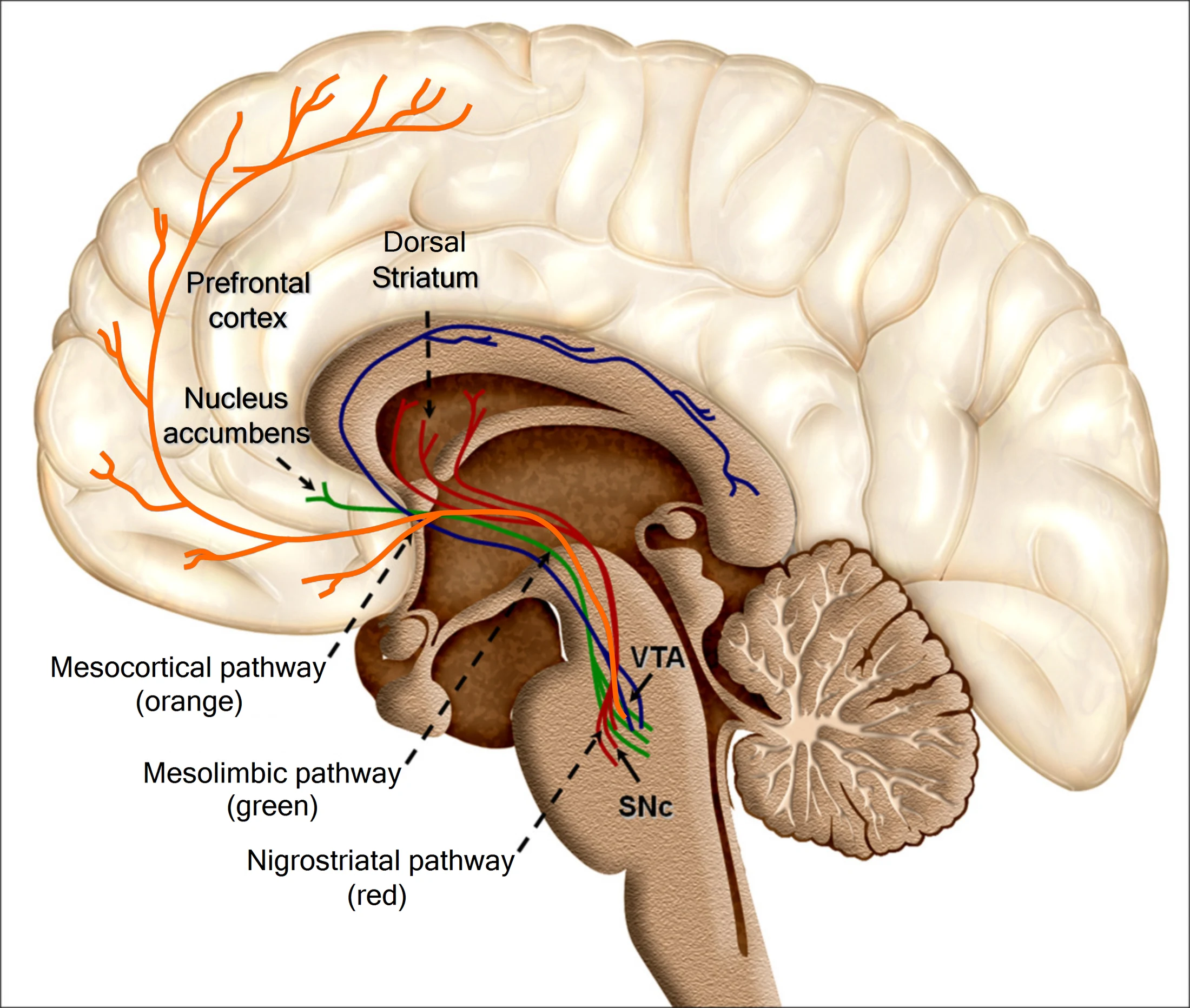
In rats, ibogaine reduced alcohol self-administration. When the drug was microinjected directly, the ventral tegmental area worked and the substantia nigra did not. Systemic administration raised GDNF in a midbrain region including the VTA, and injecting GDNF itself into the VTA reproduced the effect.
 Enlarge
That distinction matters beyond alcohol. The substantia nigra is the structure that degenerates in Parkinson’s disease, and it is the one the effect did not appear in. Ibogaine for Parkinson’s follows that thread to the two human trials of GDNF itself, both of which moved the brain imaging and failed to move the patients.
The cerebellum, where the story stops being about benefit
The third region is not a mechanism of action. It is a lesion.
In 1993 two papers reported that ibogaine destroys a subset of Purkinje cells in the cerebellar vermis of rats, preceded by activated astrocytes and microglia in the same places.
The pattern is the striking part. The damage is not diffuse. It falls in narrow parasagittal stripes within the vermis, following the cerebellum’s own longitudinal organisation, which is why the effect was recognised as selective rather than as general toxicity.
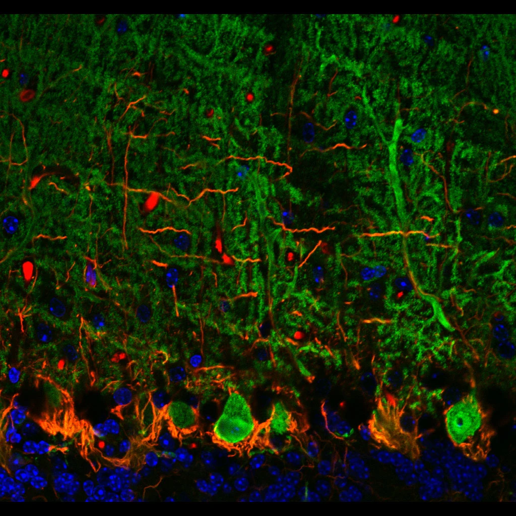

One more detail from that paper is worth carrying. Harmaline produced the same pattern. Harmaline is a beta-carboline from Banisteriopsis caapi, and it is the reason the ayahuasca comparison is not as distant as it looks. Two alkaloids from unrelated plants damage the same narrow stripes of the same structure.
The clearest picture in the field is not in the brain
Everything above is receptor densities, microinjections and rodents.
The one interaction that has been resolved to the level of individual atoms is the hERG potassium channel, and it is in the heart. How ibogaine works sets the five brain candidates against that single settled mechanism, and what it does to a heart explains why that asymmetry is the practical fact rather than a philosophical one.
What this page does not establish
Regional injection studies show that a structure is sufficient to produce an effect in an animal. They do not show that it is what happens when a person swallows the drug, and they do not show that the same circuits carry the same weight in a human brain.
There is imaging in people, and it comes from one cohort of thirty scanned by one team, described by its own authors as exploratory and never independently repeated.
So the honest summary is that the anatomy is real, mostly rodent, mostly produced with an analogue, and contradictory in at least one place that matters. What we do not know keeps the running list.
Common questions
Sources
5 sources · How we source
- Brain regions mediating α3β4 nicotinic antagonist effects of 18-MC on nicotine self-administration
- Glial cell line-derived neurotrophic factor mediates the desirable actions of the anti-addiction drug ibogaine against alcohol consumption
- Degeneration of Purkinje cells in parasagittal zones of the cerebellar vermis after treatment with ibogaine or harmaline
- α3β4 nicotinic acetylcholine receptors in the medial habenula modulate the mesolimbic dopaminergic response to acute nicotine
- Ibogaine induces glial activation in parasagittal zones of the cerebellum